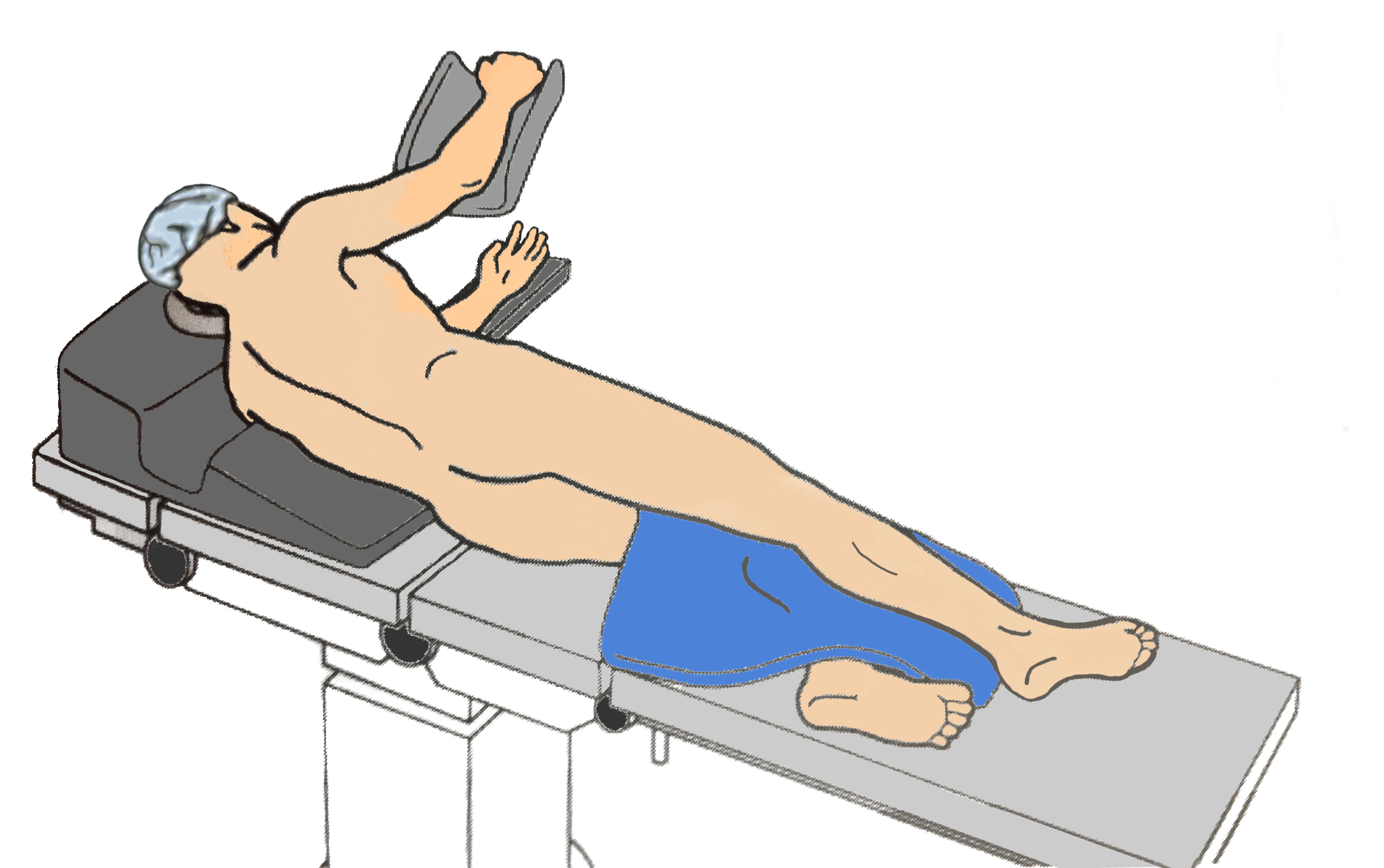
The Lateral Decubitus position is a side-lying position that provides the greatest surgical access to the thoracic organs, mediastinal contents, retroperitoneum, vertebrae, hips [1][2]. Despite optimal surgical exposure for these anatomical regions, there are a number of physiological disadvantages of the position as well as the potential for peri-operative iatrogenic injuries [1]. These physiological/respiratory changes can vary in severity based on the depth of anesthesia, use of positive pressure ventilation, and underlying co-morbidities [1]
TL;DR: Lateral Decubitus is laying on side so surgeon can get to organs, downside is respiratory changes and nerve/skin injury

Proper Positioning
Terms to Know before getting started
- Dependent: The side facing down (The Dependent side touches “D” bed)
- ABducted: bringing the limb AWAY from the body. (An ABducted child is taken AWAY from their parents)
Head
- A neutral head and neck prevents brachial plexus injury
- Careful not to fold the dependent ear
- Avoid causing pressure to the dependent eye
source: [2][3]
Upper Extremities
- Both arms are placed in front of the patient
- Both arms should be ABducted LESS THAN 90 degrees to prevent brachial plexus injury. Double check with every reposition.
- An Axillary Roll should be placed just caudal to the axilla to prevent compression of the brachial plexus and the axillary vasculature. NOTE: Despite the name the axillary roll does not go INSIDE the axilla (arm pit), this would cause direct compression of these structures and defeat the purpose…. BELOW THE AXILLA !
- The NON-dependent arm is placed on a suspended armrest or table with flexion at the shoulder and slight flexion at the elbow.
- The DEPENDENT arm is also flexed at the shoulder, and slightly at the elbow and placed in front of the patient on an arm board with padding.
- All bony prominences should be padded
- Arterial lines should be placed in the DEPENDENT arm to alert to compression of axillary vasculature
- Secure the patient to the table with retaining straps, tape, or a specialized vacuum deflatable “bean bag.”
source: [2][3][4]
Lower Extremities
- Padding placed between knees and below DEPENDENT knee
- DEPENDENT knee is slightly flexed
source: [2][3]
Respiratory Changes
Most of the respiratory changes associated with assuming the Lateral Decubitus position are due to pressure from the mediastinal contents laterally, and abdominal contents from below [1][2]. The weight of the abdominal contents result in a cephalad shift of the diaphragm on the dependent side and compliance also decreases [1][3]. The overall effects on respiration and the ventilation-perfusion (V/Q) relationship is dependent upon the patient’s level of anesthesia, as well as the use of positive pressure ventilation and/or neuromuscular blockade [1].
The Awake Patient
In the awake, spontaneously ventilating (SV) patient, perfusion is greatest in the dependent lung due to the effects of gravity on pulmonary blood flow [1]. Fortunately, the dependent lung is also the better ventilated, due to the dependent lung having more efficient hemi-diaphragmatic contraction AND a more beneficial location on the compliance curve [1]. This close matching of ventilation and perfusion in the dependent lung results in maintenance of gas exchange.
| Awake Patient Lung | Ventilation | Perfusion |
| Non-Dependent (Up) | Lower | Lower |
| Dependent Lung (Down) | Higher | Higher |
| NET RESULT: No change in V/Q |
The Spontaneously Ventilating Anesthetized Patient
Induction of general anesthesia results in a reduction of Functional Residual Capacity (FRC), with a greater decrease in the dependent lung (recall the cephalad shift of the diaphragm from abdominal contents primarily affecting the dependent side) and decreased compliance [1]. This results in inferior ventilation in the dependent lung, despite its still having better perfusion, yielding a V/Q mismatch.
| Anesthetized Patient, SV | Ventilation | Perfusion |
| Non-Dependent Lung (Up) | Higher | Lower |
| Dependent Lung (Down) | Lower | Higher |
| NET RESULT: V/Q Mismatch |
Mechanically Ventilated, Anesthetized, Paralyzed Patient
Compliance of the dependent lung is decreased due to paralysis of the hemi-diaphragm allowing abdominal contents to shift even farther cephalad, restricting ventilation of the lung [1]. This decrease in compliance can be further exacerbated by the use of the “bean bag” that aids in lateral positioning; its rigid surface prevents expansion of the dependent chest wall [1]. Positive Pressure ventilation will seek the path of least resistance, which in this case, in the much more compliant non-dependent lung, especially [1][4]. With surgical opening of the thorax on the non-dependent side there is an even greater increase in compliance, and therefore, even higher compliance relative to the dependent lung [1]. All of this, of course, results in a V/Q mismatch, considering perfusion is still superior in the dependent lung.
| Anesthetized,Ventilated, Paralyzed Patient | Ventilation | Perfusion |
| Non-Dependent Lung (Up) | Higher | Lower |
| Dependent Lung (Down) | Lower | Higher |
| NET RESULT: V/Q Mismatch |
References
[1] Butterworth IV J.F., & Mackey D.C., & Wasnick J.D.(Eds.), (2022). Morgan & Mikhail’s Clinical Anesthesiology, 7e. McGraw Hill. https://accessmedicine.mhmedical.com/content.aspx?bookid=3194§ionid=266516824
[2] Armstrong M, Moore RA. Anatomy, Patient Positioning. [Updated 2022 Oct 31]. In: StatPearls [Internet]. Treasure Island (FL): StatPearls Publishing; 2022 Jan-. Available from: https://www.ncbi.nlm.nih.gov/books/NBK513320/
[3] Gropper, M. A., & Miller, R. D. (2020). Miller’s anesthesia. Elsevier.
[4] Barash, P. G., Cullen, B. F., Stoelting, R. K., Cahalan, M. K., Stock, M. C., Ortega, R. A., Sharar, S. R., & Holt, N. F. (2017). Clinical anesthesia. Wolters Kluwer.
[5] Elisha, S., Heiner, J., & Nagelhout, J. J. (2023). Nurse anesthesia. Elsevier.
Leave a Reply